Providers turn to AI to improve scans
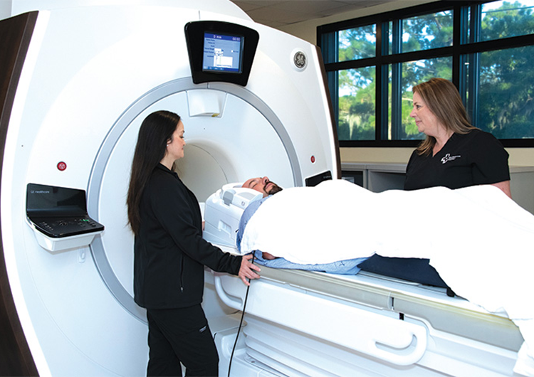
Wilmington Health Radiology’s new MRI scanner includes AirRecon DL and Artist Lift technology. (Photo courtesy of Wilmington Health)
With deep-learning algorithms and AI, health providers are starting to use a newer technology with their MRI scans on patients.
Wilmington Health Radiology has begun using a tool intended to help radiologists get sharper images. The practice’s MRI scanner now is using AIR Recon DL software to improve image resolution and clarity without increasing scan time.
“Air Recon DL is a pioneering reconstruction technology that enables our doctors to achieve sharp images faster,” said Brad Nelson, Wilmington Health’s director of radiology. “By removing noise from raw images, scans are consistently crisp. This improved technology means scan times are cut by up to 50%, and the feet-first imaging option reduces claustrophobia rejection rate by 90%.”
GE Healthcare launched AIR Recon DL, “a deep-learning image reconstruction technology, [that] makes full use of all the raw data coming off the MRI scanner,” according to the company.
In magnetic resonance, researchers have looked for ways to improve the detailed pictures, particularly in removing image noise or artifacts.
Both can make it more difficult to read accurate images of what is going on inside a patient’s body, and different approaches and technologies are used to minimize those impacts.
One way to improve the image is to collect more raw data, but that can mean more time scanning.
But in 2020 and 2021, the Food and Drug Administration gave GE Healthcare clearance to use its AIR Recon DL for reconstructing MR (magnetic resonance) images on its MRI systems. The software uses “over 100,000 unique pattern recognitions for noise and low resolution to reconstruct only the ideal object image,” according to GE Healthcare.
Last fall, the FDA cleared AIR Recon DL technology to be used with 3D imaging sequences as well.
Wilmington Health has incorporated the tool for its MRI scanner at the practice’s 1202 Medical Center Drive location. They began using the new software March 27, scanning more than 1,000 patients with it for multiple areas – including patients’ brains, bodies, vascular systems and hearts.
“We currently scan in one plane (coronal), but with this upgraded software, we have the ability to reconstruct images in all three planes, producing a 3D image,” said Holly Gillenwater, Wilmington Health’s radiology supervisor. “We already do this for specific sequences requested by the radiologist: Brain, Carotids, MRA Head. This is a huge time-saver since we only have to scan in one plane but can reconstruct in the other two.”
Matt Janik, a Wilmington Health cardiologist, uses the tool to look at cardiac MRI scans without needing additional radiology readings.
“The AI technology is an engineering breakthrough that allows coil design that is lighter, offers more flexibility and provides greater coverage,” he said, referring to MRI machine coils that essentially act as antennas transmitting radio frequency signals from patients’ bodies to computers to create the images.
“Along with enhancing workflow and the patient experience,” Janik added, “the Artist Lift Technology is the most cost-effective way to gain the benefits of a next-generation MRI system.”